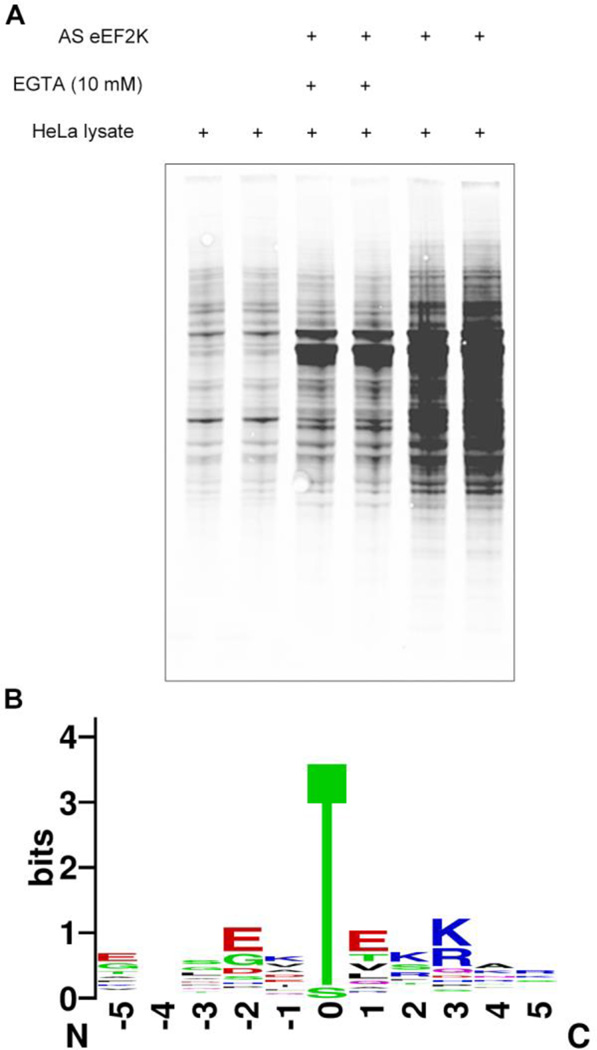
Figure 2. Discovery of substrates of eEF2K.
(A) Phosphorylation of substrates in HeLa lysate was detected using the thiophosphate adduct antibody. Each reaction was performed in duplicate. A small amount of each reaction was removed for this Western blot analysis. The rest was captured on resin for mass spectrometry analysis. (B) Sequence motif for substrates detected in mass spectrometry experiments selectively with eEF2K and calcium. Position 0 shows a strong preference for threonine. Motif was generated using WebLogo(47)

