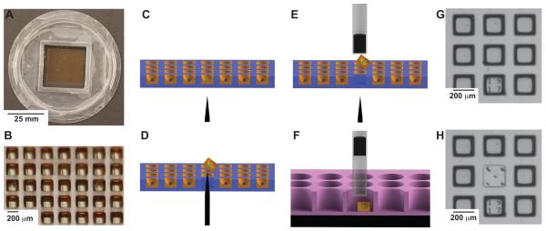
Figure 1. Microraft based selection of cells.
The microraft array in these studies is a 2.5 cm × 2.5 cm array containing 70 × 70 microrafts. Each microraft is 200 μm × 200 μm × 200 μm in outer dimensions and separated from adjacent microrafts by a 100 μm gap. The cassette walls surrounding the array are 1 cm high allowing for excess media to be applied over the microrafts (Figure 1A, B). Fluorescence from each microraft can be measured. In this example a “green cell” is identified (Figure 1C). The array is encapsulated in gelatin, and the needle release mechanism pushes through the PDMS to release the identified microraft (Figure 1D). The magnetic wand attached to the microscope objective captures the released microraft (Figure 1E). The wand is moved to a well of a 96-well plate where a block magnet pulls the microraft to the bottom of the well (Figure 1F). A 3 × 3 section of the array is shown using bright field microscopy (Figure 1G). The needle release ejected the microraft in the middle. In this example 5 needle penetrations were used to demonstrate the accuracy of the device and to show that the elastomeric PDMS readily reseals without loss of media despite repeated puncturing (Figure 1H).

