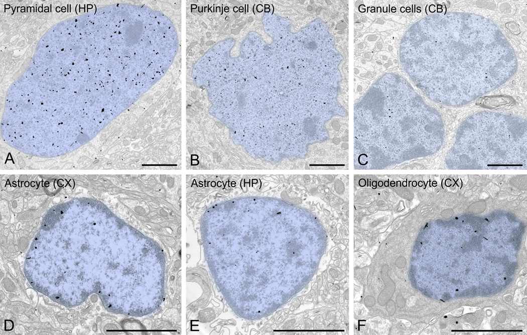
Figure 10.
UBE3A in neuronal and glial nuclei. Label in neuronal nuclei (A, CA1 pyramidal neuron; B, Purkinje cell; C, cerebellar granule cells) is denser than in glial nuclei (D, astrocyte from cerebral cortex; E, astrocyte from CA1 hippocampus; F, oligodendrocyte from cerebral cortex), with distinct patterns of distribution. The neuronal label tends to avoid heterochromatin electron-dense zones, whereas label in glia tends to associate with heterochromatin. Scale bars = 2 µm.

