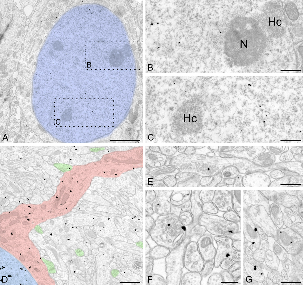
Figure 8.
Pre-embedding immunogold labeling for UBE3A in cerebral cortex at P7. A–C: The pattern of nuclear labeling is similar to that seen in the adult (compare with Fig. 4); label avoids heterochromatin patches and the nucleolus. D: General view shows label in nucleus (colorized blue) and proximal dendritic shaft (pink). Some label can be seen in multiple compartments of neuropil, including axon terminals (green). E–F: Label concentrates over developing vesicle-rich axon terminals. G: Label can be seen at the edge of mitochondria. Scale bars = 2 µm in A; 500 nm in B, C; 1 µm in D; 500 nm in E; 250 nm in F; 500 nm in G.

