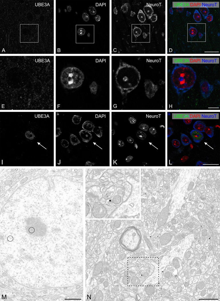
Figure 9.
UBE3A staining in the primary visual cortex of adult AS mice model. A–L: High magnification immunofluorescence, showing UBE3A staining in cerebral cortex (primary visual cortex, layer I-II). Sections were counterstained with DAPI (red) to visualize nuclei, and with NeuroTrace 640–660 (blue) to visualize neuronal somata. UBE3A staining is confined to weakly immunoreactive puncta in the neuropil. Rare neurons exhibit nuclear staining similar to that seen in WT mice (arrows, in I–L).
M, N: Pre-embedding immunogold labeling for UBE3A in cerebral cortex (primary visual cortex, layer I–II). Only very rare silver/gold particles are located over neuronal nuclei (circles in M). In the neuropil, labeling can be seen in presynaptic terminals (inset in N). Scale bars = 20 µm in A–D and I–L; 5 µm in E–H; 1 µm in M, N.

