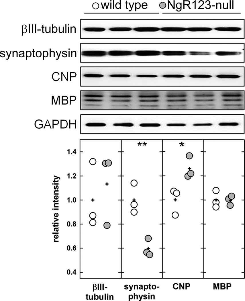
Figure 3.
Immunoblots of neuronal, synaptic and myelin markers in extracts of midline structures from wild type and NgR123-null mice. Homogenates of midline structures dissected from 3 mice of each group were resolved by SDS-PAGE, transferred, and blotted with specific antibodies for neurons (anti-βIII-tubulin), synapses (anti-synaptophysin), myelin (anti-CNPase and anti-MBP) and a loading control protein (anti-GAPDH). Densitometry of each band was normalized to GAPDH in the same sample. Normalized data (relative to GAPDH) were compared by Student’s t-test (*, p < 0.05; **, p < 0.01). Data points are presented relative to the wild type average, with each data point for wild type (open symbols) and NgR123-null (grey symbols) presented. Averages for each genotype are denoted with a plus sign (+).

