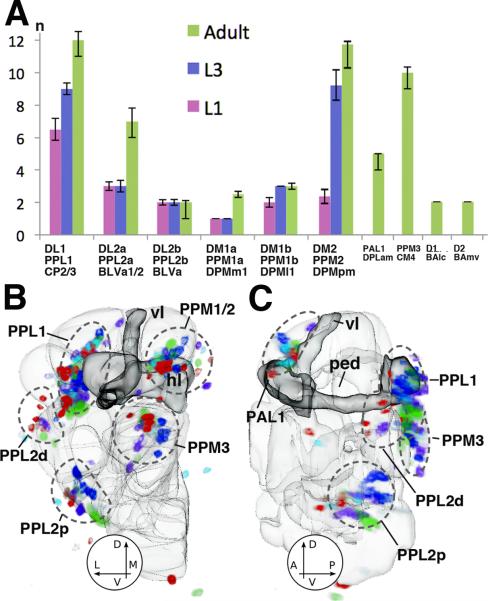
Fig.3.
Cell numbers and variability of Drosophila DA neurons. (A) Plot of cell numbers per one brain hemisphere for different clusters of DA neurons at early larval stage (L1), late larval stage (L3), and adult (n=5). (B, C) Variability in number and position of DA neurons in the adult brain. Both panels show 3D digital models of left brain hemisphere in posterior view (A) and lateral view (B). Outlines of compartments (light gray) and mushroom body (dark gray) are shown as reference. Labeled cell bodies of DA neurons of five registered brain hemispheres are shown in different colors. One color (e.g., red) represents all DA neurons of one brain hemisphere. The overlay of five brains still allows to recognize the main clusters of DA neurons (PPL1, PPL2d/p, PPM1-3); however, there is considerable scatter of somata within clusters. Abbreviations of compartments: ML medial lobes of the mushroom body; PED peduncle; VL vertical lobes of the mushroom body.

