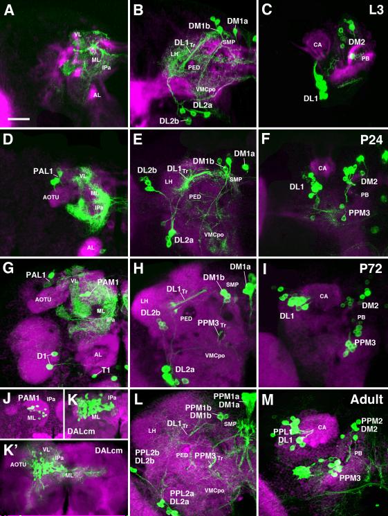
Fig.4.
Development of pattern of DA neurons from larva to adult. All panels present z-projections of frontal confocal sections of brain hemisphere at late third instar larval stage (A-C), 24h pupa (D-F), 72h pupa (G-I), and adult (J-M). Dorsal up, medial to the right. DA neurons are labeled with TH-Gal4 > UAS-mCD8::GFP (green). Secondary lineages are labeled by anti-Neurotactin (red in A-C) or anti-Neuroglian (red in D-I, L, M). Panels of the left column (A D, G, J-K’) represent anterior slice of the brain at the level of the mushroom body lobes (ML medial lobe; VL vertical lobe). Middle column (B, E, H, L) shows posterior slice at level right behind central complex; right column (C, F, I, M) represents z-projections far posterior, at level of calyx (CA) and protocerebral bridge (PB). Clusters of DA neurons, located in the anterior brain (left column) or posterior brain (middle and right column) are annotated. Individual clusters can be followed throughout metamorphosis based on location and axonal trajectory. For adult (bottom row), both larval and adult annotation is provided (e.g. PPL2b/DL2b). Note DA clusters that become TH-positive during metamorphosis: PPM3 and PAL1, corresponding to the lineages CM4 and DPLam, respectively, are recognizable from 24h onward (D, F); PAM1 first differentiates in the 72h pupa (G, J-K’). Also note strong reduction of terminal arborizations of DA neurons during pupa (E, H), compared to larva (B) or adult (L). Other abbreviations: AL antennal lobe; AOTU anterior optic tubercle; CA calyx; IPa anterior domain of inferior protocerebrum; LH lateral horn; ML medial lobe; PED peduncle; PB protocerebral bridge; SMP superior medial protocerebrum; VL vertical lobe; VLP ventrolateral protocerebrum; VMCpo posterior domain of ventromedial cerebrum. Bar: 25μm

