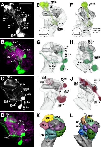
Fig.6.
Terminal arborization of DA neurons in early larval brain. A-D: Z-projections of confocal sections of L1 brain hemisphere. DA neurons are labeled by TH-Gal4 > UAS-mCD8::GFP (white in A, C; green in B, D). In B and D, neuropil compartments are visualized by anti-DN-cadherin (magenta). Plane of confocal sections is tilted at a 45deg angle relative to the horizontal plane, as shown by the line in panel F. E-L: 3D digital models of first instar larval brain hemisphere. Models of left column (E, G, I, K) show anterior view (dorsal up, medial to the left); right column presents lateral view (dorsal up, anterior to the left). In E-J, neuropil compartments are shown semitransparent and in gray color; compartments are rendered in different colors in panels K and L. E-J present volume renderings of the three most conspicuous DA clusters of the early larva in different colors (E, F: DM1 light green; G, H: DL2 dark green; I, J: DL1 magenta). In all panels, DA clusters are annotated in a manner that points out the location of somata (so), axon tract (ax), and terminal arborization (term). Abbreviations of compartments: AL antennal lobe; CA calyx of mushroom body; VL vertical lobe of mushroom body; IPa anterior domain of inferior protocerebrum; IPl lateral domain of inferior protocerebrum; IPm medial domain of inferior protocerebrum; CPLd primordium of superior lateral protocerebrum and lateral horn; CPM primordium of central complex and posterior inferior protocerebrum; LAL lateral accessory lobe; ML medial lobe of mushroom body; PED peduncle; PENP periesophageal neuropil; SEG subesophageal ganglion; SMP superior medial protocerebrum; SP spur; VLP ventrolateral protocerebrum; VMC ventromedial cerebrum; Bar: 20μm

