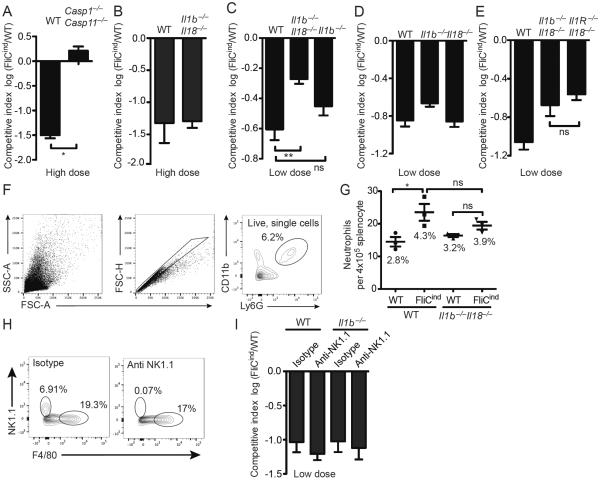
Figure 1. IL-1β, IL-18, and pyroptosis cooperatively clear FliCind S. Typhimurium.
(A–E, H, I) Mice (n=5) were infected with (A, B) high dose (105 CFUs) or (C–I) low dose (103 CFUs) (of 1:1 ratio of FliCind AmpR and WT KanR S. Typhimurium IP for 17 hours prior to doxycycline injection; organs were harvested 7 hours later. CIs are expressed as the log FliCind/WT (Amp/Kan CFUs). (F–G) Mice were infected with WT or FliCind S. Typhimurium and treated with doxycycline at 24 hours pi for 5 hours. Spleens were prepared for single cell suspension, stained and analyzed by flow cytometry. Neutrophils were defined as the CD11b+ Ly6G+. (F) The gating strategy represents a WT mouse infected with FliCind S. Typhimurium. (G) The mean percentage of neutrophils of the total splenocytes population is stated in the figure for each condition. (H–I) For NK-cell depletion, mice were injected with 75 ng anti-NK1.1 PK136 or isotype control C1.18.4 (BioXCell) at −3 and −1 dpi. Depletion of NK1.1-positive cells was confirmed by flow cytometry (left). NK cells were defined as NK1.1+ F4/80−, and the percentage of each cell type is indicated. All data are shown as mean + SE of n=3 (F, G) and n=5 (A–E, H, I) mice and are from single experiments representative of two (A, H, I) or three (B–G) experiments. * p < 0.05; two-tailed student t-test.

