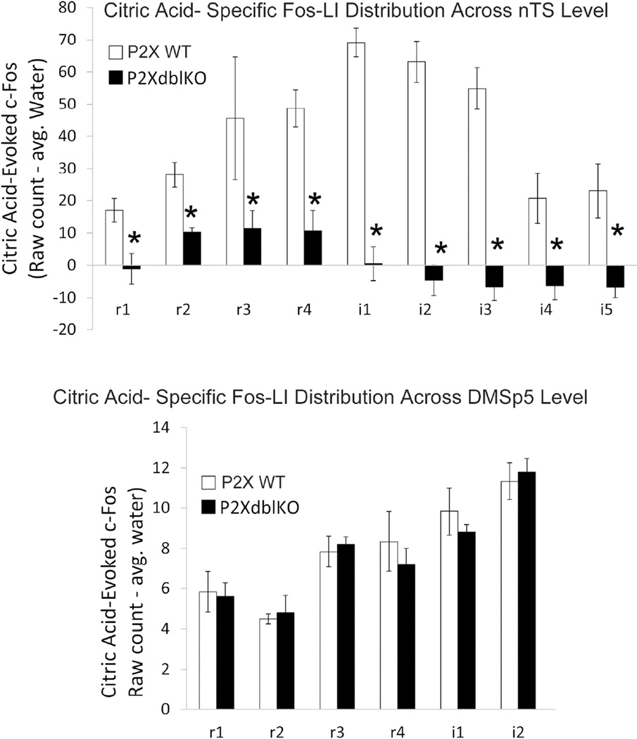
Figure 12. Quantitative comparison of citric acid-evoked Fos-LI (raw count – avg. water) within the brainstem of P2X WT and P2X-dblKO mice across the different levels of the nTS and in the DMSp5.
A. The number of Fos-LI neurons is significantly reduced across all nTS levels in the nTS of P2X-dbl KO mice, (* = Post hoc Tukey’s HSD results from main Group×nTS level ANOVA interaction is p < 0.05). Note also the paucity of Fos-LI in P2X-dblKO mice in more caudal portions of the nTS (i4, i5) wherein esophageal afferents terminate. B. In contrast, Fos-LI is similar between P2X WT and P2X-dbl KO mice in the dorsomedial trigeminal nucleus (DMSp5).

