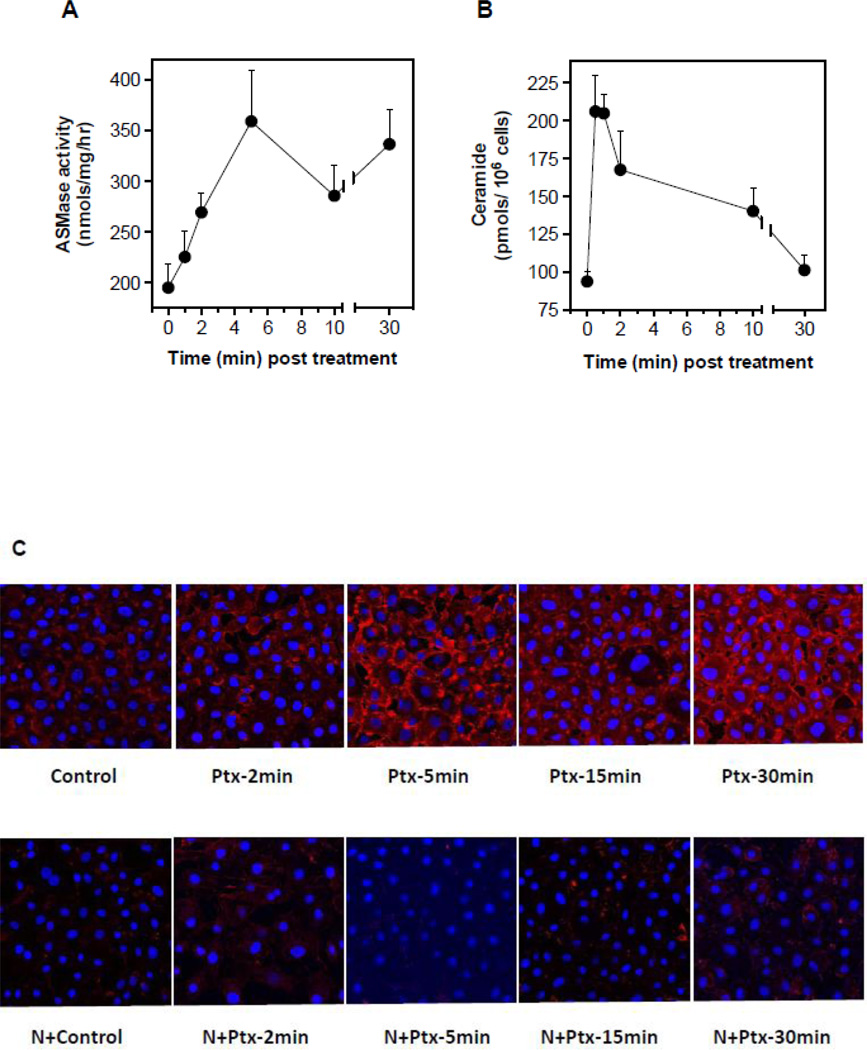
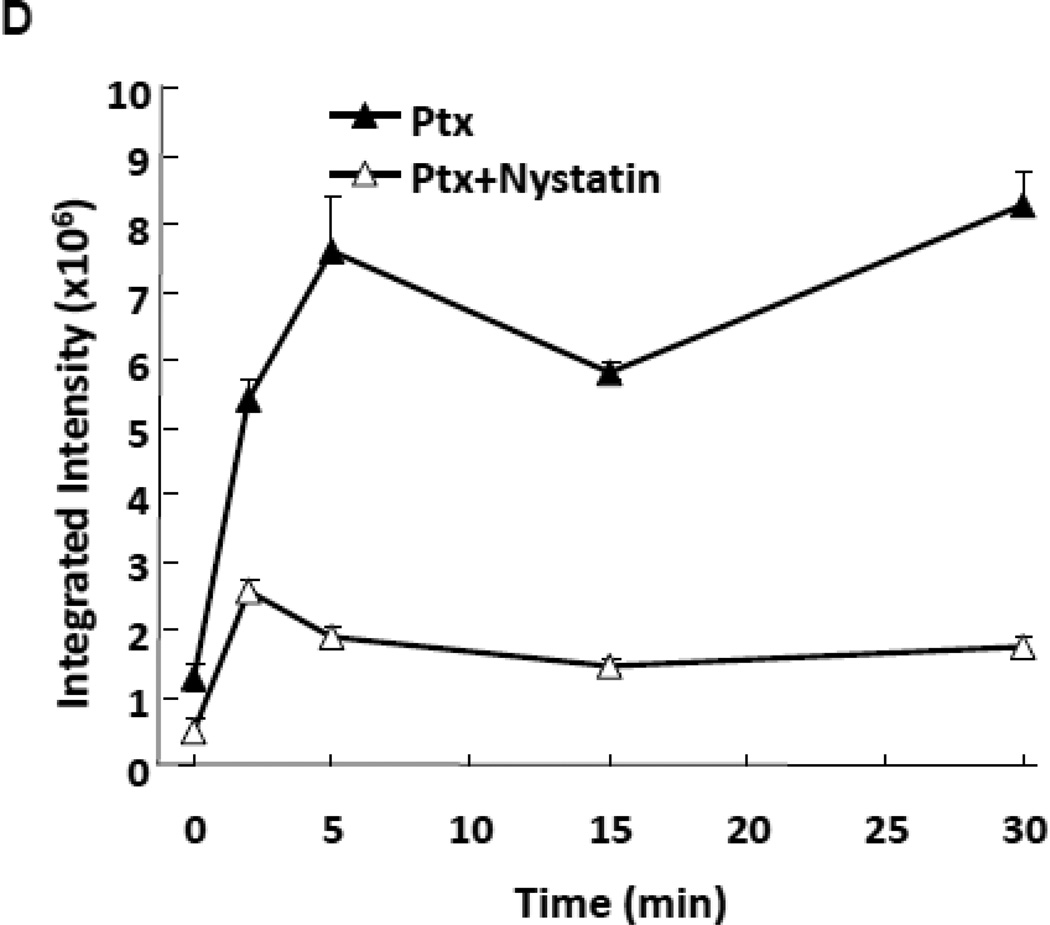
Figure 1.
ASMase activation and CRMs generation by paclitaxel in cultured endothelium. (A) ASMase activity was quantified in lysates of BAEC at the indicated times after treatment with paclitaxel (100 nM) by radioenzymatic assay using [N-methyl-14C] sphingomyelin as substrate. (B) In parallel, a time-course of ceramide generation was measured using the diacylglycerol kinase assay. (C) BAEC monolayers, exposed to paclitaxel (100 nM) in the absence (upper panel) or presence (lower panel) of 30 µg/mL nystatin (30 min pre-treatment), were co-stained with anti-ceramide antibody (red) and DAPI (blue, to stain nuclei) in order to localize CRMs to plasma membranes by confocal microscopy. (D) The integrated intensity of the CRMs = the sum of the ceramide intensity (above background) multiplied by the area for each CRM. Data (mean±SD) represent triplicate determinations from duplicate experiments each in A, B and D.


