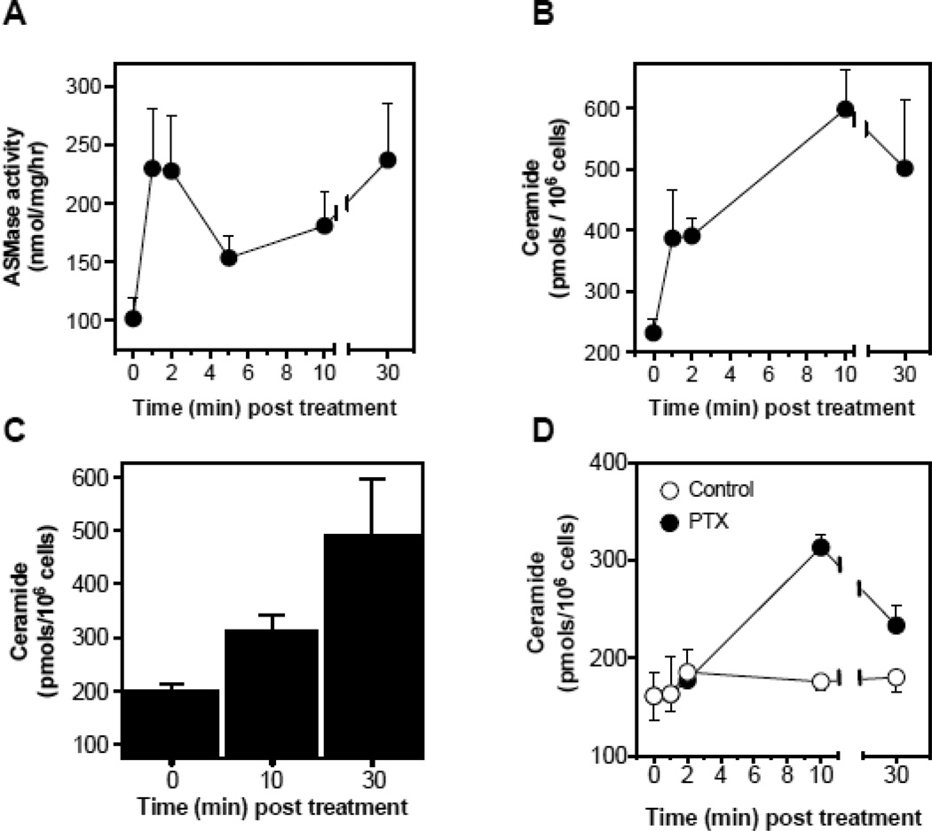
Figure 2.
Chemotherapy-induced ASMase activation, ceramide generation and induction of apoptosis in cultured endothelium. (A) In a time-course experiment, ASMase activity was quantified in BAEC after treatment with 50 µM etoposide by radioenzymatic assay using [N-methyl-14C] sphingomyelin as substrate. (B) In parallel, a time-course of ceramide generation in response to etoposide was measured using the diacylglycerol kinase assay in BAEC. (C) Ceramide generation was measured using the diacylglycerol kinase assay at 10 and 30 min after treatment in HCAEC with 50 µM etoposide. (D) Ceramide generation was measured using the diacylglycerol kinase assay at 10 and 30 min after treatment in HCAEC with 100 nM paclitaxel. Data (mean±SD) represent triplicate points from two independent experiments.

