Figure 3.
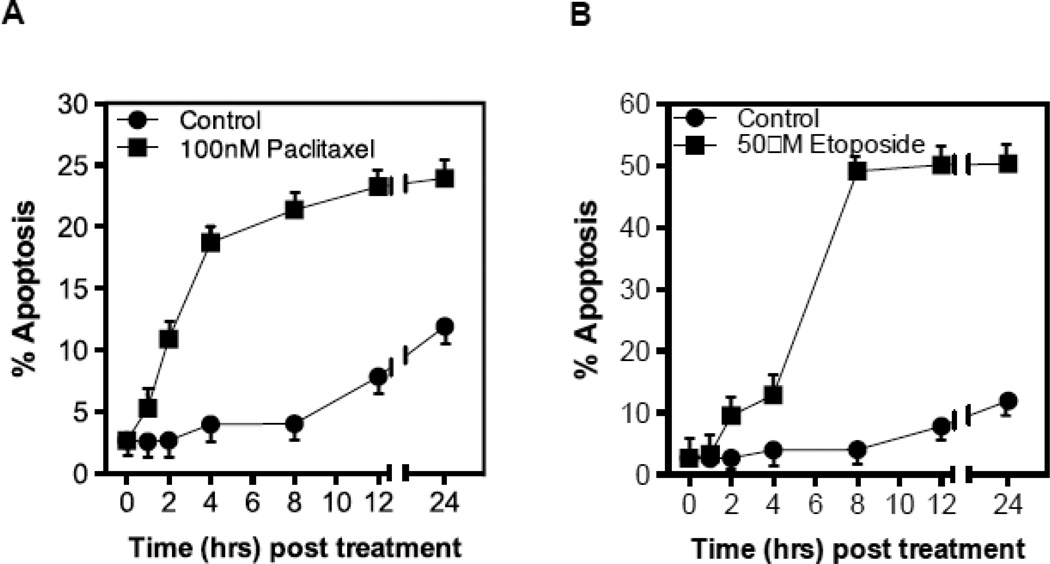
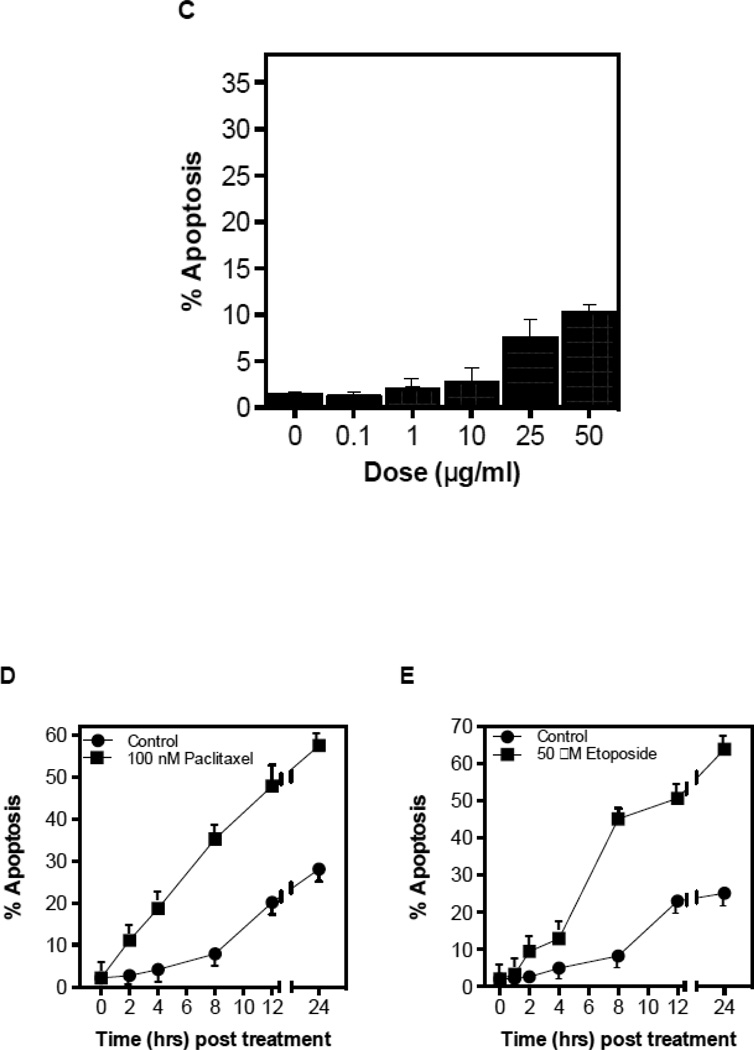
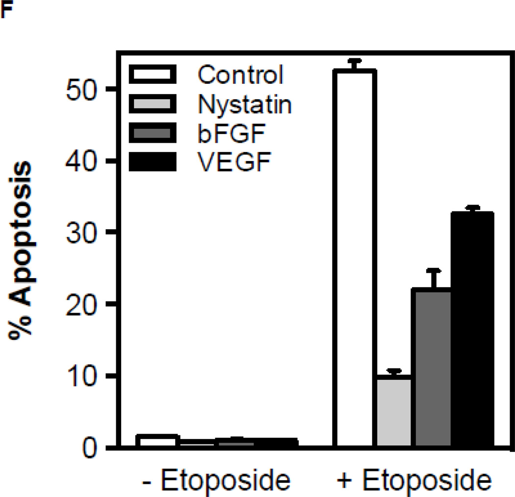
CRM generation signals chemotherapy-induced apoptosis in endothelium. Cultured BAEC were treated with paclitaxel (100 nM) (A) or etoposide (50 µM) (B) and at the indicated times incidence of apoptosis was scored by bis-benzamide trihydrochloride staining. (C) Treatment of BAEC with cisplatin does not lead to endothelial cell dysfunction. BAEC were treated with increasing doses of cisplatin (0.1–50 µM) and apoptosis was detected by bis-benzamide trihydrochloride staining. Paclitaxel and etoposide induce apoptosis in HCAEC. HCAEC were treated with 100 nM paclitaxel (D) or 50 µM etoposide (E) and the incidence of apoptosis was scored at the time points indicated. (F) BAEC were pre-incubated for 30 min with bFGF (2 ng/mL), VEGF (2 ng/mL) or nystatin (30 µg/mL) prior to treatment with etoposide (50 µM), and apoptosis was evaluated after 8 hours. Each value (mean±SD) represents duplicate determinations from three independent experiments.


