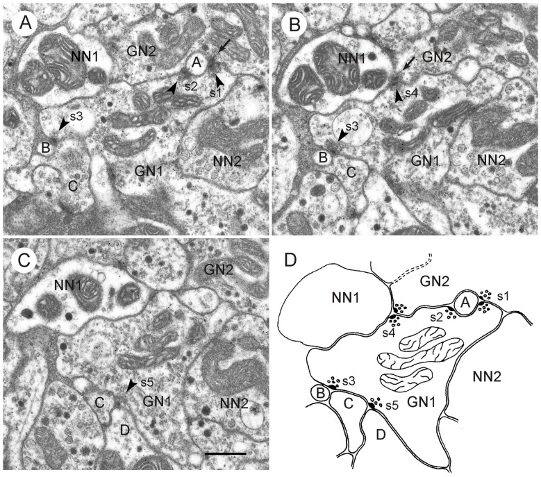
Figure 5.
GABA-immunoreactive neuron with output synapses. (A–C) Details from section three (A), five (B) and eight (C) out of eight consecutive immunogold-labeled sections through the CBL. The GABA-immunoreactive profile (GN1) makes five output synapses (s1-s5) to neighboring profiles (A–D, NN1). Arrowheads point to presynaptic densities. Two of these synapses (s1 in A and s4 in B) are of type II, i.e., the presynaptic profile (GN1) and a second presynaptic profile (GN2) face one postsynaptic profile (A and NN1). GN2 is like GN1 GABA-immunolabeled. The postsynaptic profile NN1 of s4 was not immunolabeled; it contains a few dense core vesicles. Whether profile A is GABA-immunolabeled or not, could not be determined. Output synapses s2, s3 and s5 are of type I. In synaptic profile s2, (A) GN1 is presynaptic to profile A and to the second GABA-immunolabeled profile GN2. In synaptic profiles s3 (A,B) and s5 (C) GN1 is presynaptic to profiles B and C (s3), resp. profiles C and D (s5). To the right of GN1, an unlabeled profile (NN2) contains granular vesicles. The mean gold particle (GP) densities (n = 8) were 13.0 ± 4.1 GP/μm2 (GN1), 8.0 ± 2.5 GP/μm2 (GN2) 1.8 ± 1.4 GP/μm2 (NN1), and 1.2 ± 0.9 GP/μm2 (NN2). The GP densities in GN1 and GN2 were significantly different from those in NN1 and NN2 (one-way ANOVA, Tukey-HSD, p < 0.05). (D) Schematic diagram illustrating synaptic contacts of GN1 in the eight sections. Scale bar in (C): 0.5 μm (applies to A–D).

