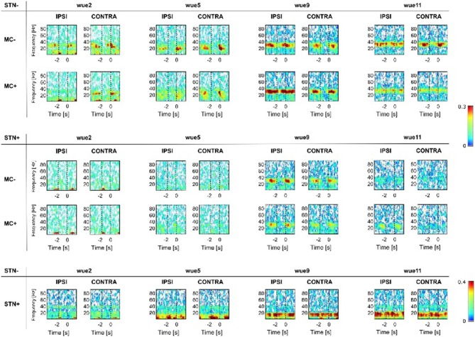
Figure 2.
Coherence analyses. Subcortical- (i.e., CohSTN−/STN+) and cortical-subcortical coherence (i.e., CohSTN−/MC−, CohSTN−/MC+ and CohSTN+/MC−, CohSTN+/MC+) are reported for each subject with respect to STN− and STN+. White color shows lack of coherence. From blue to red color we show increasing significant coherence between brain structures. Results were mirrored by iCoh, thus supporting the lack of volume conduction artifact (Supplementary Material, Results, Figure S8). As in Figure 1, the super-imposed vertical dotted line at 0 s shows the RT. We also indicated, with a dotted line at −1.7 s, the mean OT of all trials (see also Figure S1) as a rough indication of movement OT. “IPSI” and “CONTRA” refer to movement performed with the hand ipsilateral or contralateral to the examined STN (STN− or STN+). “−” (MC− and STN−) and “+” (MC+ and STN+) refer instead to the side with less and more striatal dopaminergic innervation or the more and less clinically affected hemibody (for wue05). MC, motor cortex; STN, subthalamic nucleus. MC, motor cortex; STN, subthalamic nucleus.

