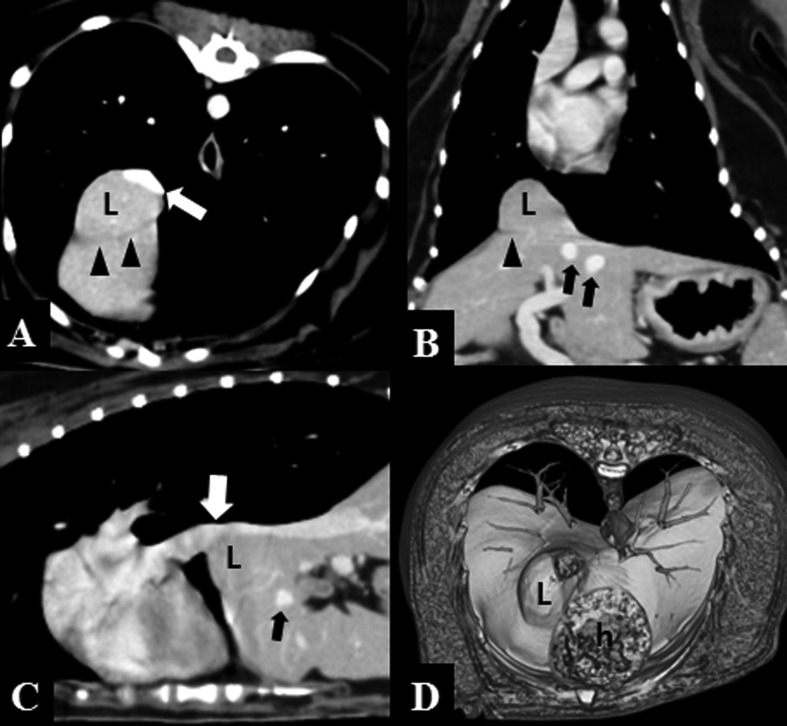
Fig. 2.
Post-contrast CT images of a typical caval foramen liver hernia in dog 3. Partial herniation of the right lateral liver lobe (L) at the central tendon of the diaphragm compresses the adjacent caudal vena cava dorsally (white arrows). Dilation of the main hepatic veins (black arrows) and the intact muscular portion of diaphragm (arrowheads) are visible. (A, transverse plane; B, dorsal plane; C, sagittal plane; D, volume-rendered image, diaphragmatic aspect; h, heart)

