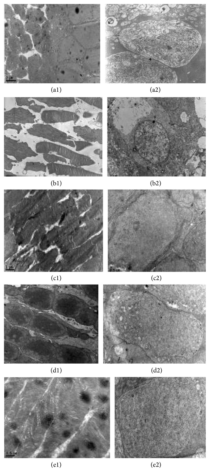
Figure 3.

Ultrastructure of retina. (a1) Structure of photoreceptor cells was clear; rod and cone cells were arranged in alignment. (a2) Ganglion cells were round or ovoid, with obvious nuclei. Inside they were full of organelles as mitochondria, rough endoplasmic reticulum, golgi apparatus, and so forth. (b1) Part of photoreceptor cells was rupture, with tumid and vacuolar degenerative mitochondria. Rod outer segments had fuzzy skyline. (b2) Ganglion cells decreased in numbers and in microfilament and microtubules components. Organelles such as mitochondria, rough endoplasmic reticulum, or golgi apparatus almost disappeared. ((c1), (d1)) Arrangement of photoreceptor cells was mildly disordered. There were no vacuolar degenerative mitochondria. ((c2), (d2)) Ganglion cells decreased in numbers, but in the nuclei there was homogeneous chromatin. Organelles such as mitochondria, rough endoplasmic reticulum, or golgi apparatus could be seen with mild degeneration. (e1) Structure of photoreceptor cells was distinct; rod and cone cells arranged in alignment, without obvious degeneration. (e2) Ganglion cells were nearly normal in structure. Inside there were clear organelles as mitochondria, rough endoplasmic reticulum, golgi apparatus, and so forth.
