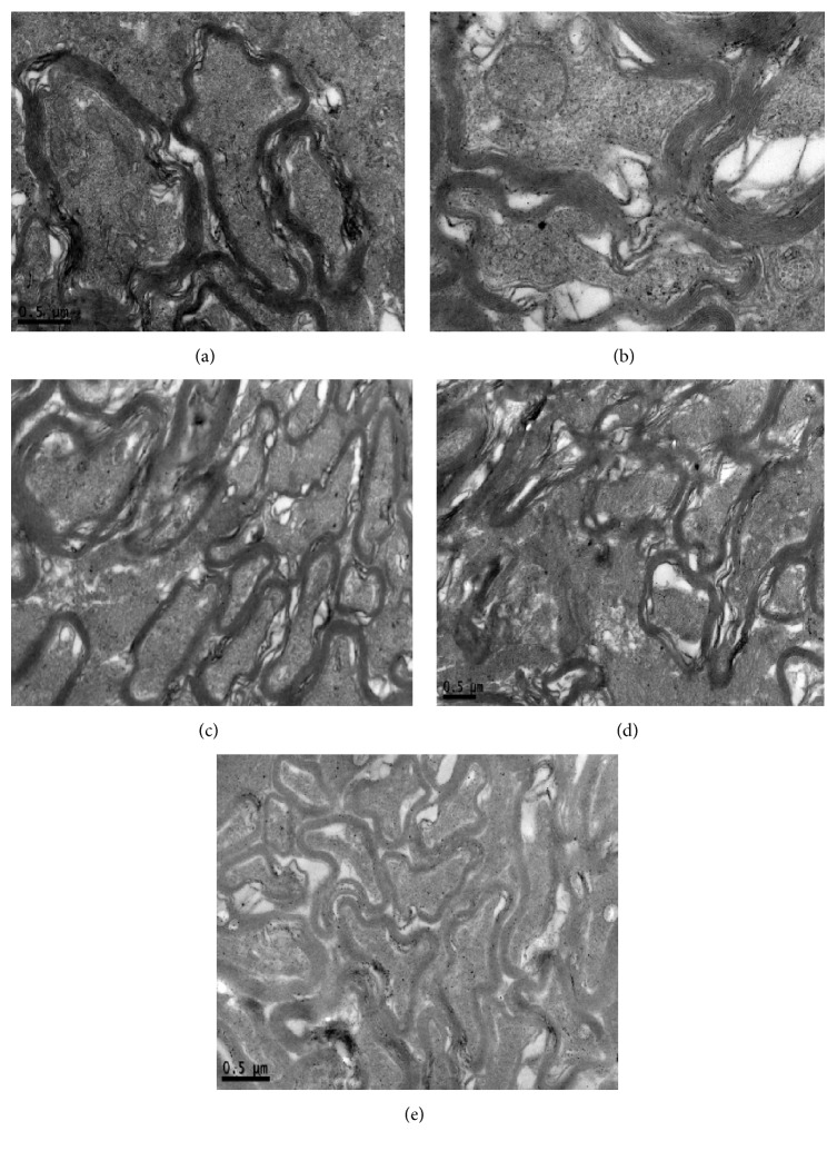
Figure 4.
Ultrastructure of optic nerve. (a) The structure of myelin sheath was complete. In the axoplasm, microtubules, microfilaments, and organelles such as mitochondria could be seen explicitly. (b) The dissolved myelin sheath was loose. In the axoplasm, microtubules and microfilaments became swallowing. Vacuolar degenerative mitochondria could be seen. (c) (d) Part of myelin sheath was attenuation. In the axoplasm, microtubules and microfilaments became mildly swallowing. Vacuolar degenerative mitochondria could also be seen. (e) The structure of myelin sheath was complete but fair-arranged. In the axoplasm, microtubules, microfilaments, and organelles such as mitochondria could be seen without degeneration.

