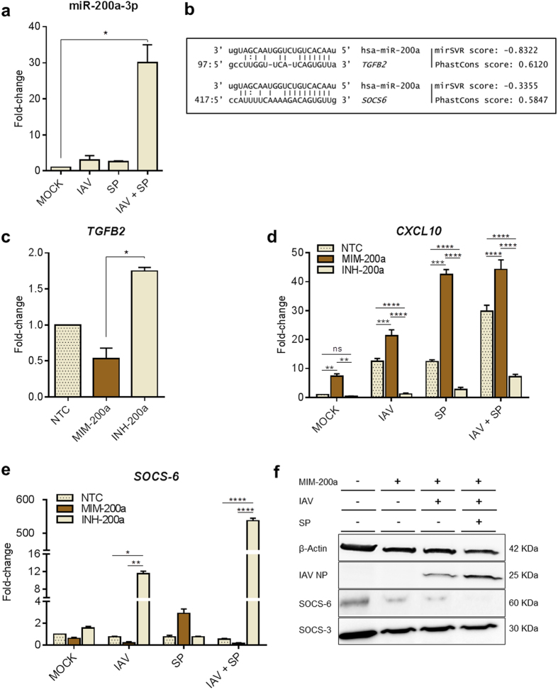
Figure 3. Functional analysis of the synergistic induction of miR-200a-3p in MDMs after mixed infection with IAV and SP.
(a) Preceding IAV infection elevates miR-200a-3p expression after mixed infection with SP. MDMs were infected with IAV for 4 hours, then infected with SP, incubated for 20 h and total endogenous miRNAs were purified. A specific PCR-probe assay targeting miR-200a-3p was used to assess the fold changes in miR-200a-3p expression in mock-transfected and infected cells. (b) In silico predictive target alignment showing that miR-200a-3p targets the 3′ UTRs of both TGFB2 and SOCS6. (c) TGFB2, (d) CXCL10 and (e) SOCS6 expression profiles in MDMs transfected with negative transfection control (NTC), miR-200a mimic (MIM-200a) or miR-200a inhibitor (INH-200a). At 18 h after transfection, the MDMs were singly or mixed infected as described previously. At 8 h post-IAV and/or SP infection, total mRNA was extracted and amplified by PCR using specific primers for the indicated genes. Values represent median ± IQR (a, c) or mean ± SEM (d, e) of three biological replicates. Statistical analyses were performed using a Kruskal-Wallis test (non-parametric, one-way ANOVA with Dunn’s post-hoc test) for data presented in (a, c). An ordinary two-way ANOVA (with Tukey’s post-hoc multiple comparison test) was used for data presented in (d, e). *P < 0.05; **P < 0.01; ***P < 0.001; ****P < 0.0001. (f) Western blot analysis of SOCS-6, SOCS-3, IAV nucleoprotein (NP) and β-actin expression in MDMs transfected with negative transfection control (NTC), miR-200a mimic (MIM-200a) or miR-200a inhibitor (INH-200a), cultured for 18 h, and then infected as described previously. Cell lysates were harvested 24 hours post-infection.

