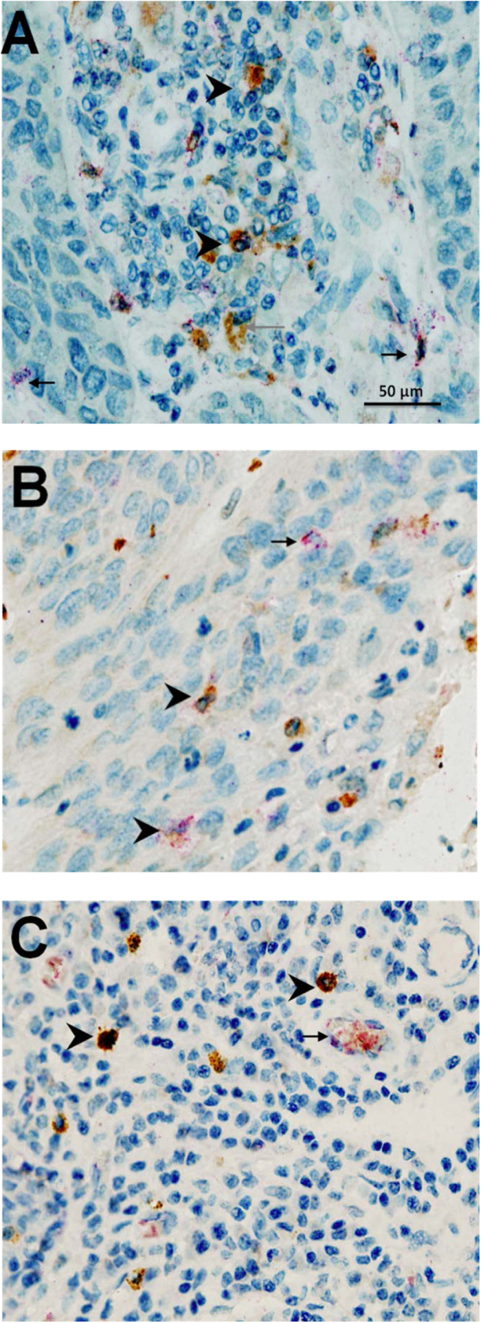
Figure 1.

Examples of immunohistochemical double-staining for (A) chymase (brown) and tryptase (red) demonstrating the presence of MCT mast cells (red) and MCTC (reddish brown) (B) tryptase (brown) and TNFα (red) demonstrating the presence of TNFα in tryptase + mast cells, and (C) chymase (brown) and TNFα (red) demonstrating the expression of TNFα in MCTC mast cells. Arrowhead = double-stain cell. Black arrow = single-stain red cell. Grey arrow = single-stain brown cell.
