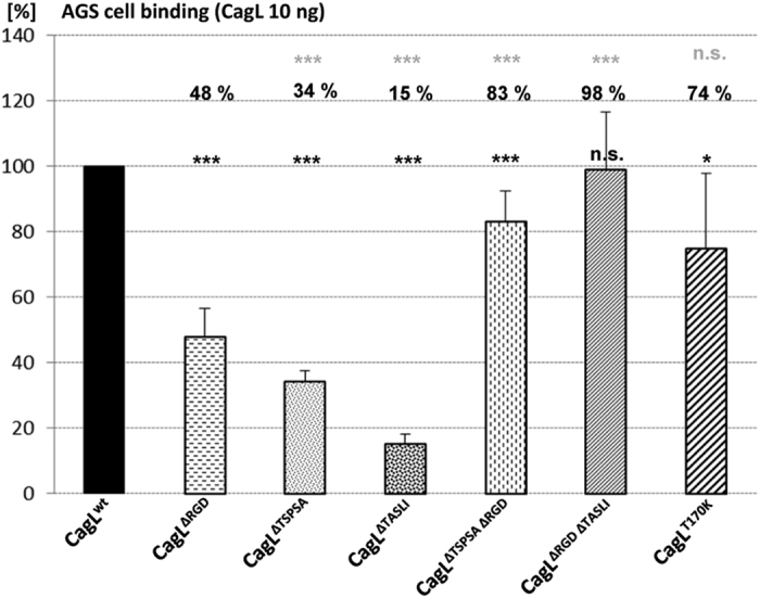
Figure 6. Differential binding of site-directed CagL mutants to AGS human gastric epithelial cells.

Highly purified native CagL variants (ng amounts, as indicated) were incubated in microwell plates containing fixed adherent AGS cells for 3 h, followed by extensive washing, and quantitated by an ELISA-like method using anti-CagL antibody (Methods). The results are depicted as relative values in comparison to the CagL wild type control, which was set to 100%. Cell binding by different CagL mutants (10 ng each of highly pure protein) in comparison to wild type CagL is shown. Significances of differences to CagL wild type binding were calculated by student’s t-test (two-tailed, unpaired) from the mean values and SDs of three independently performed experiments, summarizing six data points in total, and are shown as black asterisks above each bar: ***p < 0.01; *p < 0.05. Upper row of asterisks: significance of differences between CagLΔRGD mutant and other mutants, calculated on the basis of the same experiments. n.s. = not significant.
