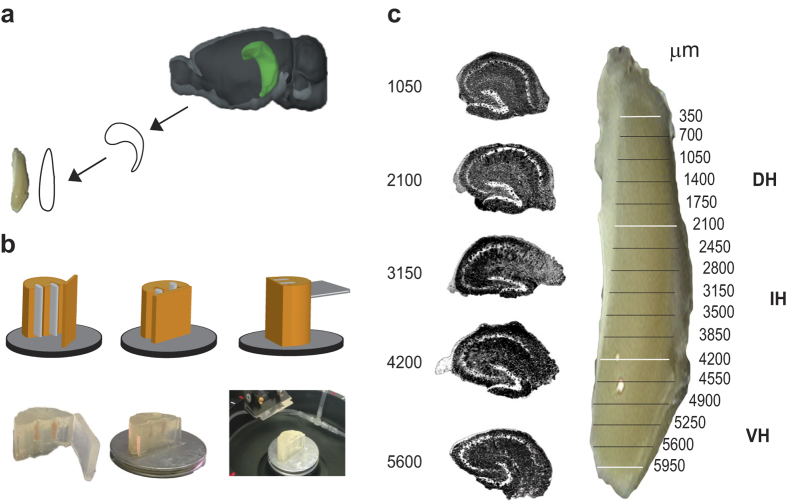
Figure 1. Hippocampal slicing procedure.
(a) Schematic diagram showing isolation and elongation of the hippocampus. Image credit: © 2015 Allen Institute for Brain Science. Allen Brain Atlas API. (b) The hippocampus is placed in a small agarose-block fixed to the cutting stage of the vibratome magnetic plate in order to prepare 350 μm thick transverse slices. (c) Schematic partition of the hippocampus along the septo-temporal axis and representative Hoechst- stained slices at different septo-temporal positions distances. White lines indicate boundaries among different zones, the black ones indicate the longitudinal position of slices used for electrophysiological recording.

