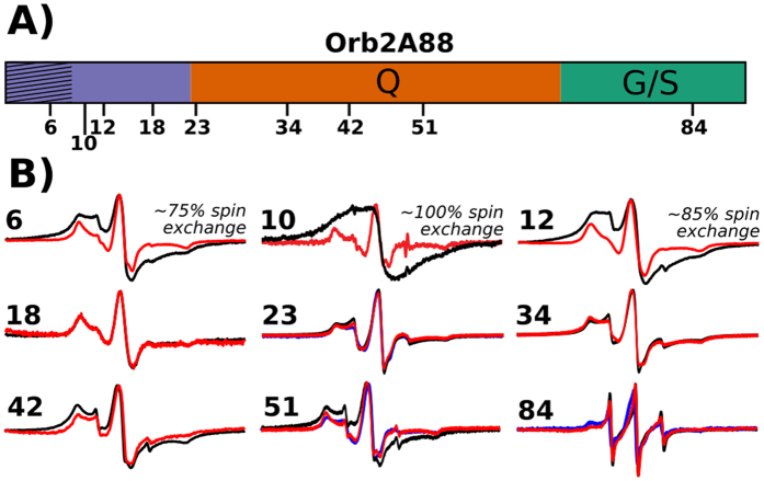
Figure 5. EPR spectra show that the N-terminal amyloid core of Orb2A88 is in an in-register parallel β-sheet conformation.
(A) Domain structure of Orb2A88 showing positions that were spin labeled. (B) EPR spectra of Orb2A88 fibrils spin labeled at the indicated positions. 100% MTSL labeled spectra are shown in black. 10% MTSL labeled samples that were diluted with an MTSL analogue are shown in red. As an additional control we made 10% MTSL labeled spectra of residue 23, 51, and 84 that were diluted with the Cys free C10M mutant instead (shown in blue). Spectra are shown at same amplitude. The percentage of spin exchange calculated from comparing 10% with 100% labeled spectra is given when present. The single EPR line seen for the 100% labeled sample in contrast to the 10% labeled samples is indicative of spin exchange often observed in parallel, in-register β-sheet amyloids22.

