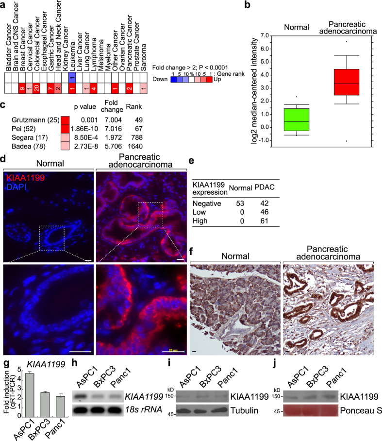
Figure 2. Expression of KIAA1199 in human pancreatic ductal adenocarcinoma (PDAC).
(a) Expression of KIAA1199 in human cancers via Oncomine analysis. (b,c) Expression of KIAA1199 in human pancreatic adenocarcinoma via Oncomine analysis. (d–f) Expression of KIAA1199 in human pancreatic adenocarcinoma. Fluorescence immunohistochemical analysis of KIAA1199 of human pancreatic cancer tissue microarray (TMA; US Biomax PA207), using anti-KIAA1199 antibody (d). Quantitative analysis of KIAA1199 expression in TMA (e). Chromogenic immunohistochemical analysis of human pancreatic cancer tissue microarray (US Biomax BIC14011a, PA242b) using-KIAA1199 antibody and DAB substrate (f). (g,h) mRNA expression of KIAA1199 in PDAC cell lines (AsPC-1, BxPC-3, and Panc-1). Quantitative RT-PCR (g) and semi-quantitative RT-PCR (h). KIAA1199 expression in PDAC cell lines compared to HPNE (normal pancreas epithelial cell line) and normalized to 18 S rRNA. Gel images shown have been cropped to show the relevant band. Full-length gels are presented in Supplementary Figure 3. (i,j) Endogenous and secreted protein expression of KIAA1199 in PDAC cell lines (AsPC-1, BxPC-3, and Panc-1). Molecular weight of KIAA1199 was approximately 150 kDa. Blot images shown have been cropped to show the relevant band. Full-length blots are presented in Supplementary Figure 3.

