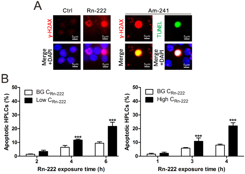
Figure 5. Apoptotic pan-nuclear γ-H2AX response in HPBLs in vitro exposed to radon and its progeny.
(A) Representative images of pan-nuclear γ-H2AX staining in HPBLs induced by in vitro exposure to radon and its progeny and the co-localization of pan-nuclear γ-H2AX signal with TUNEL staining in HPBLs induced by α-particle irradiation from 241Am source at 6 h post-irradiation. (B) Relationship between the percentages of pan-nuclear γ-H2AX-positive cells and radon exposure time in HPBLs. One thousand HPBLs from each sample were used for quantitation. The data are presented as the mean ± standard deviation of three human blood samples. Red, γ-H2AX; Green, TUNEL staining; blue, DNA stained with DAPI. 1000 × magnification. ***P < 0.001 compared with the control group that was exposed to the natural background levels of radon. BG, background.

