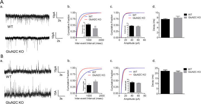
Figure 1. Opposing alterations in excitatory and inhibitory activity of layer V pyramidal neurons in the mPFC of GluN2C KO mice.
(A) A significant reduction was observed in the frequency, but not amplitude or decay, of mEPSCs of layer V pyramidal neurons in the mPFC of GluN2C KO mice. Representative traces (a); Quantitative data (n = 20 for WT and 13 for KO from 4–5 mice/genotype) cumulative probability plots of inter-event interval and frequency in inset, significant reduction in mEPSC frequency in GluN2C KO was observed (unpaired t-test with Welch’s correction, **P = 0.0035) (b); cumulative probability plot for amplitude and mean amplitude in inset (c) and decay (d) of mEPSCs. (B) A significant increase was observed in the frequency, but not amplitude or decay of mIPSCs from layer V pyramidal neurons in the mPFC of GluN2C KO mice. Representative traces (a); Quantitative data (n = 11 for WT and 16 for KO from 4–5 mice/genotype) cumulative probability plots of inter-event interval and frequency in the inset, significant increase in frequency of mIPSC in GluN2 KO was observed (unpaired t-test with Welch’s correction, *P = 0.0204); cumulative plot for amplitude and mean amplitude in inset (c); and decay (d) of mIPSCs.

