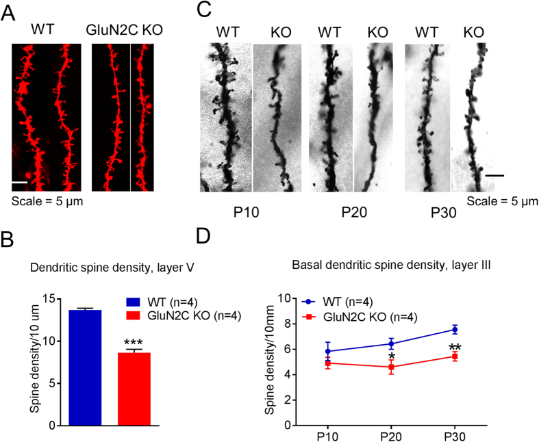
Figure 2. Lower dendritic spine density in medial prefrontal cortex of GluN2C knockout mice.
(A,B) Dendritic spine density of pyramidal neurons in layer V of the medial prefrontal cortex (mPFC) was evaluated using diolistic labeling and Imaris analysis. (A) Representative images and (B) quantitative results. A significantly lower total spine number was observed in GluN2C KO compared to WT (n = 4 mice/genotype, total of 81 neurons in WT and 76 neurons from GluN2C KO; unpaired t-test, ***P = 0.0002). (C,D) Basal dendritic spine density of pyramidal neurons in layer III of the mPFC was determined using Golgi staining. (C) Representative images of Golgi-stained sections and (D) quantitative results. Basal dendritic spine density was significantly lower in GluN2C KO mice at P30 (n = 4/genotype, unpaired t-test with Welch’s correction, **P = 0.0061 < 0.01) and P20 (n = 4/genotype, unpaired t-test with Welch’s correction, *P = 0.0447), with a trend towards reduction at P10.

