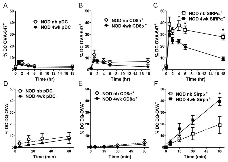
Figure 4. 4 wk-old thymic SIRPα+ DC exhibit enhanced antigen processing and presentation properties.
Percent of OVA-647+ thymic (A) pDC, (B) CD8α+, and (C) SIRPα+ from newborn or 4 wk-old NOD mice injected with 2.7 mg/kg (3.5 μg or 50 μg respectively) of OVA-647 (n≥ 8 mice/group) at different times post injection. (D–F) Frequency of fluorescent thymic DC from newborn and 4 wk-old NOD mice treated with 0.5 μg/well DQ-OVA in vitro. Data ± SEM are the average of 4 separate experiments. *p<0.05 (A–C, Student’s t-test; D–F, linear regression analysis).

