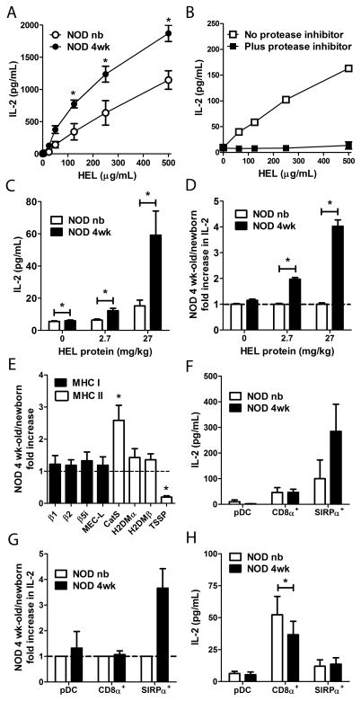
Figure 5. 4 wk-old SIRPα+ DC demonstrate enhanced Ag processing and T cell stimulation.
Newborn and 4 wk-old NOD thymic DC (A–E) in bulk or (F–H) flow-sorted subsets were tested for inducing IL-2 secretion by (A–D, F, G) Clone 4 CD4+ or (H) 8c11 CD8+ HEL-specific T cells. (A,B, F–H) Thymic DC were pulsed with (A,B) varying concentrations, (F,G) 125 μg/ml, or (H) 500 μg/ml of HEL protein in vitro, or (C,D) isolated from newborn and 4 wk-old NOD mice injected i.v. with 2.7 or 27 mg/kg body weight of HEL, respectively. (B) IL-2 induced secretion following pre-treatment of NOD 4 wk-old thymic DC with a protease inhibitor cocktail. (D,G) Fold increase in IL-2 production induced by 4 wk-old versus newborn thymic DC with the newborn values equal to 1. (E) Relative expression of MHC I and II protein processing molecules in NOD newborn and 4 wk-old thymic DC determined by real-time PCR. (H) Sorted thymic DC subsets were co-cultured with 500 μg/mL HEL protein plus Clone 8c11 CD8+ T cells and IL-2 measured. Data ± SEM of n=2 (B,F,G), n=3 (A,C,D,H), n=4–5 (E) separate experiments are depicted. *p<0.05 (Student’s paired t-test).

