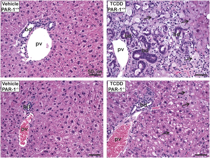
FIG. 4.
Light photomicrographs of hematoxylin and eosin (H&E)-stained liver tissue sections from male mice gavaged with sesame oil vehicle or 30 µg/kg TCDD every 4 days for 28 days. Marked bile duct (bd) hyperplasia, hepatocellular vacuolization (arrows), and periportal inflammatory cell infiltration in the liver is shown. Pv indicates the portal vein. Scale bars represent 50 µm.

