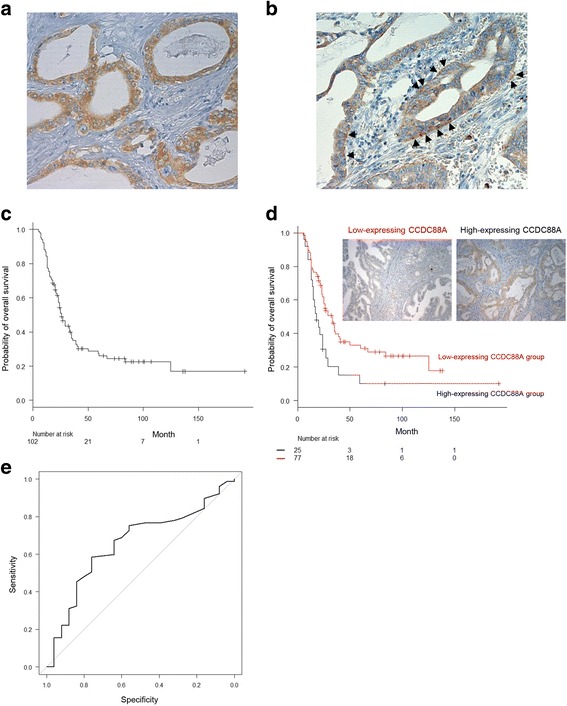
Fig. 1.

Association of high expression of CCDC88A with a poor outcome in PDAC patients. a, b. Immunohistochemical staining of PDAC tissues using anti-CCDC88A antibody. CCDC88A staining was present in the cytoplasm (a) and in the basolateral portions (b) of tumor cells. Arrows, CCDC88A localized basolaterally. Magnifications: ×200. c, d. Kaplan-Meier analysis of (c) PDAC-specific survival and (d) overall survival according to CCDC88A expression. e. ROC curve of 102 PDAC cases for analysis of the impact of the total immunohistochemical score of CCDC88A on prognosis
