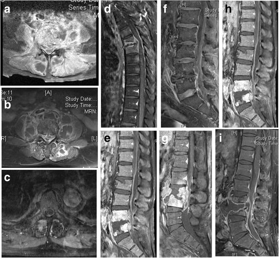
Fig. 3.

Elucidation of epidural abscess and muscle abscess in contrast-enhanced magnetic resonance imaging with fat suppression among 9 g-positive pyogenic spondylodiscitis (a–c). The spinal epidural abscess was defined as an epidural mass with iso-or hypointensity on T1-weighted images, which was surrounded by linear enhancement (ring sign) on magnetic resonance imaging. Abscess formation in psoas muscle or back muscle was defined as an asymmetrical enlarged mass of the involved muscle with ring sign on magnetic resonance imaging. d–f Ventral epidural abscess was noticed on sagittal gadolinium-enhanced fat-suppressed T1-weighted magnetic resonance
