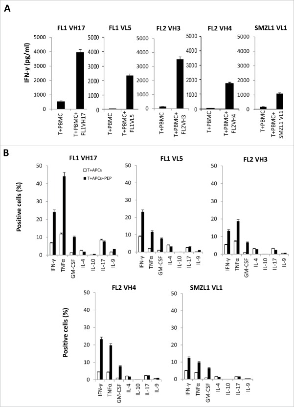
Figure 1.
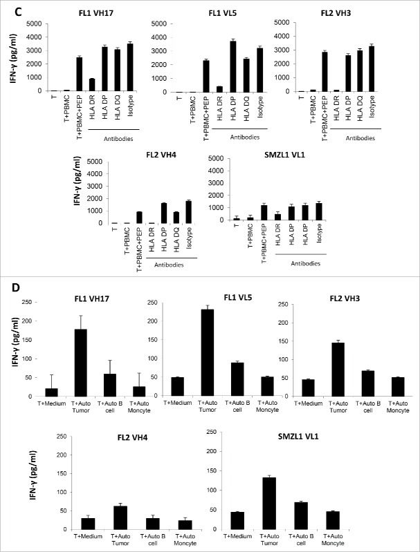
Generation of Th1 CD4+ T-cell lines against BCR overlapping peptides from autologous lymphoma patients. (A) IFNγ ELISA assay of autologous CD4+ T cells stimulated with autologous PBMCs, pulsed or nonpulsed with patient-derived 15-mer BCR overlapping peptides. Briefly, PBMCs (1 × 10⁵ cells/well) were stimulated with 10 μg/mL of each peptide in a 96-well, U-bottom-microculture plate every 3 d. After five stimulations, T cells from each well were washed and incubated with PBMCs in the presence or absence of the corresponding peptide. The production of interferon (IFN)γ was determined in the supernatants by ELISA after 18 h. (B) Intracellular cytokine staining of autologous BCR peptide-specific CD4+ T cells stimulated by APCs, pulsed or nonpulsed with peptides. (C) Blocking of IFNγ production by autologous BCR peptide-specific CD4+ T cells by HLA antibodies. (D) Recognition of autologous tumor by BCR peptide-specific CD4+ T cells. Data are representative of three individual experiments. FL, follicular lymphoma; SMZL, splenic marginal zone B-cell lymphoma.
Figure 1.
(Continued)


