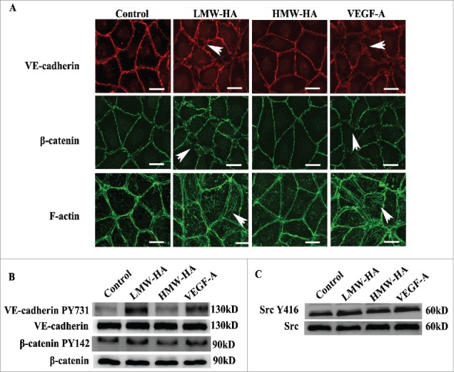
Figure 4.

Mechanism studies of the effects of LMW-HA on lymphatic intercellular adhesion. (A) Effects of LMW-HA on VE-cadherin, β-catenin and F-actin localization in HDLECs were evaluated by immunofluorescence and phalloidin staining. After treatment with LMW-HA (10 µg/mL) or VEGF-A (100 ng/mL) for 2 h, VE-cadherin (red) and β-catenin (green) became disorganized, and numerous stress fibers (green) traversing the cell body were observed (as indicated by the white arrows). Cells were visualized under a confocal microscope. One representative image of three experiments is shown. (Scale bars: 25 µm.). (B, C) The levels of VE-cadherin, β-catenin, and Src phosphorylation in HDLECs were measured. HDLECs were stimulated with 10 µg/mL of LMW-HA, 10 µg/mL of HMW-HA, 100 ng/mL of VEGF-A, or medium for 30 min. Stimulated cell lysates were subjected to immunoblot analysis with the indicated antibodies. GAPDH was the loading control.
