
Figure 5.
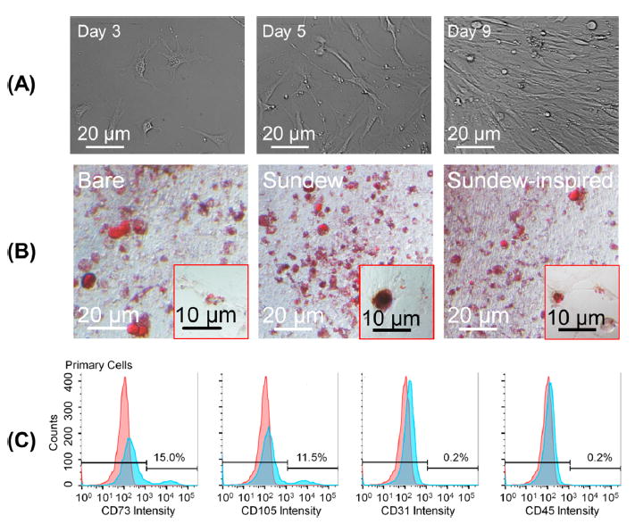
Characterization of isolated ADSCs. Morphological observations, Oil Red O stainning, and flow cytometry were used to characterize isolated ADSCs. (A) Morphological changes in isolated primary ADSCs on lysine-coated culture plates over time. The ADSCs proliferated rapidly in vitro, forming a homogeneous composition in monolayer, with fibroblastlike morphology. (B) ADSCs were plated onto bare culture plates or culture plates modified with sundew-inspired or -derived hydrogels and then induced with adipocyte differentiation medium. On day 7, cells were stained with Oil Red O. The result suggested that the sundew and sundew-inspired adhesive hydrogels do not suppress the differentiation of ADSC and thus are capable of acting as scaffolds that support the functionalities of ADSCs on wound sites. (C) Characterization of isolated ADSCs by determing expression level of CDs on the cytoplasmic membrane using flow cytometry. The ADSCs were positive for mesenchymal markers CD73 and CD105 and negative for endothelial marker CD31 and hematopoietic markers CD45, indicating that they are pluripotent.