Figure 1.

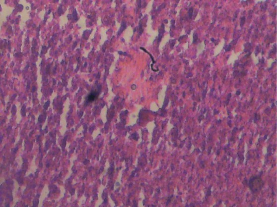
Liver section of male control rat. Cords of hepatocytes well preserved and essentially normal, cytoplasm not vacuolated, sinusoids well demarcated, no area of necrosis, no fatty degeneration and change. Haematoxylin and Eosin stained.

Liver section of male control rat. Cords of hepatocytes well preserved and essentially normal, cytoplasm not vacuolated, sinusoids well demarcated, no area of necrosis, no fatty degeneration and change. Haematoxylin and Eosin stained.