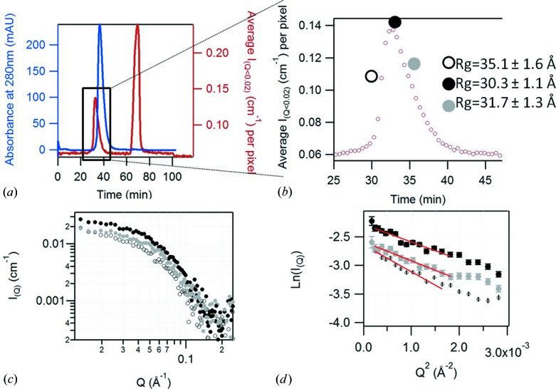
Figure 4.
(a) Results of the SEC-SANS measurement of protonated Sir2a (4 mg ml−1) in 100% D2O buffer, showing the absorbance of the sample at 280 nm (in blue) and the averaged SANS intensity between Q = 0.012 Å−1 and Q = 0.041 Å−1 (in red). (b) Magnified plot of SANS intensity of the Sir2A elution peak. The large symbols show the three individual positions, selected at the beginning (open circle, R g = 35.1 ± 1.6 Å), the top (black circle, R g = 30.3 ± 1.1 Å) and the end (gray circle, R g = 31.7 ± 1.3 Å) of the elution peak, from which are extracted individual SANS curves (c) (30 s exposure) and the Guinier plot (d) (the red lines being the fits of the Guinier region).

