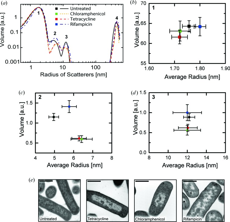
Figure 2.
Cellular composition of E. coli cells after antibiotic treatment determined by SAXS. (a) Volume distribution of the cellular components before and after antibiotic treatment as a function of scatterer’s radius. The total volume and the average radius of each scattering population were extracted from this distribution. (b) Average radius and volume of population 1, corresponding to the size range of proteins. (c) Average radius and volume of population 2, corresponding to three aggregated DNA fibers covered with histone-like proteins. (d) Average radius and volume of population 3, corresponding to ribosomes. The displayed errors bars in (b)–(d) denote the standard deviation of the model from the experimental data calculated with the uncertainty module of the IRENA toolbox. (e) Transmission electron micrographs of E. coli after antibiotic treatment. The scale bar has a length of 1 µm.

