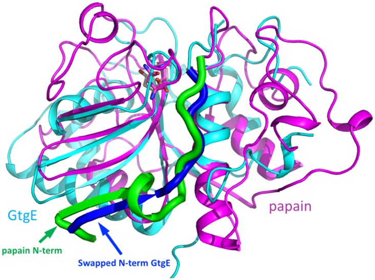
Fig 4. Superposition of GtgE (cyan) and papain (magenta). Papain structure used for the comparison has PDB code 3CVZ [33].

(A) The N-terminus of papain is shown as a thick green ribbon. The swapped N-terminus of GtgE from a neighboring molecule is shown as a thick blue ribbon. GtgE and papain superimpose well within the core of the fold, enclosed within the blue circle. (B) Close-up of the catalytic triads Cys-His-Asp/Asn in this superposition. The Ser45 of GtgE was replaced here by a cysteine and the sidechain of His151 was rotated by 180°. The catalytic residues are shown in stick representation, magenta carbons in papain, white carbons in GtgE.
