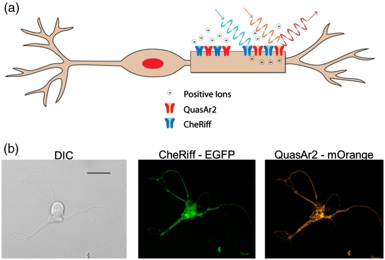
Fig. 1.
(a) Drawing of a neuron expressing the Optopatch2 proteins CheRiff and QuasAr2. CheRiff is a cation channel activated by blue light. QuasAr2 is fluorescent and the fluorescence intensity increases with membrane voltage. (b) Representative confocal differential interference contract and fluorescence images of a neuron, scale bar is . Fluorescence image of enhanced green fluorescent protein (EGFP) confirms successful transfection of CheRiff-EGFP. Fluorescence image of QuasAr2-mOrange confirms successful transfection of QuasAr2-mOrange.

