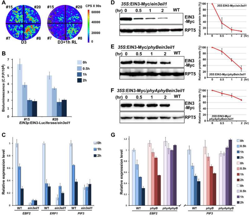
Figure 2. Red light induces rapid degradation of EIN3 through phyA/phyB to repress ethylene-responsive genes.
(A) Bioluminescent images of EIN3p-EIN3-Luciferase/ein3eil1 seedlings. Four independent transgenic lines were grown in the dark for 3 days, and then maintained in darkness (D3) or exposed to red light (D3+1h RL) for 1 hr before photographing. The color-coded bar indicates the intensity of luciferase activity. C.P.S is Counts Per Second.
(B) Quantitative analysis of EIN3-Luciferase bioluminescence with the time course of red light irradiation. Two representative lines of 3-day-old dark-grown EIN3p-EIN3-Luciferase/ein3eil seedlings were subjected to red light irradiation for the indicated times. C.P.P is Counts Per mg of Protein.
(C) qRT-PCR results showing the expression of EIN3 target genes in WT and ein3eil1. The seedlings were grown in the dark for 3 days (0h) and then subjected to red light irradiation for the indicated times. Gene transcription was normalized to PP2A. Mean ±s.d., n=3.
(D-F) Western blot results for determination of the protein levels of EIN3 upon light exposure. Seedlings over-expressing EIN3-Myc in an ein3eil1 (D), phyBein3eil1 (E) or phyAphyBein3eil1 (F) background were grown in the dark for 4 days (0 hr) and then transferred to red light irradiation for the indicated times. WT was used as a negative control. RPT5 was employed as a loading control. Right panels show the quantification analysis of three biological replicates of EIN3-Myc protein levels after normalization to RPT5. The dark EIN3-Myc protein level was set as 100%. Mean ± s.d., n=3.
(G) qRT-PCR results showing the expression of EIN3 target genes in WT, phyB and phyAphyB. Four-day-old etiolated seedlings were transferred to red light irradiation for the indicated times. Gene expressions were normalized to PP2A. Mean ± s.d., n=3.

