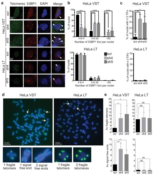
Figure 4.
Oxidized nucleotides induce telomere defects in cells with shortened telomeres. HeLa cell lines with very short telomeres (VST) or long telomeres (LT) were analyzed 3 days after tansduction with lentivirus expressing a non-targeting shRNA (scr) or different shRNAs against MTH1 (sh4 and sh5). (a) 53BP1 foci (red) were visualized by immunofluorescence and telomeric foci were detected by fluorescence in situ hybridization (green). Additional immunostaining of telomeric RAP1 protein was required in HeLa VST cells to amplify the telomere signal. 53BP1 foci at telomeres appear yellow (white arrow). (b) The number of 53BP1 foci per nuclei after binning. Bars represent the mean ± sd from 3 independent experiments (100 – 150 cells per condition). ** p<0.01, *** p<0.001, **** p<0.0001, One-way ANOVA with Tukey’s honest significance difference. (c) The percent of nuclei showing ≥ 3 telomeric 53BP1 foci per nuclei (TIFs). Bars are the mean ± s.d. from 3 independent experiments. * p<0.05, One-way ANOVA. (d) Representative metaphase chromosomes of telomere FISH from HeLa VST and HeLa LT cells expressing sh5 against MTH1. Images of fragile telomeres and signal free ends are shown. (e) Quantitation of telomere aberrations, average number per metaphase. Bars represent the mean ± sd from 3 independent experiments (at least 15–20 metaphases per condition).* p<0.05, ** p<0.01, two-tailed Student’s t test.

