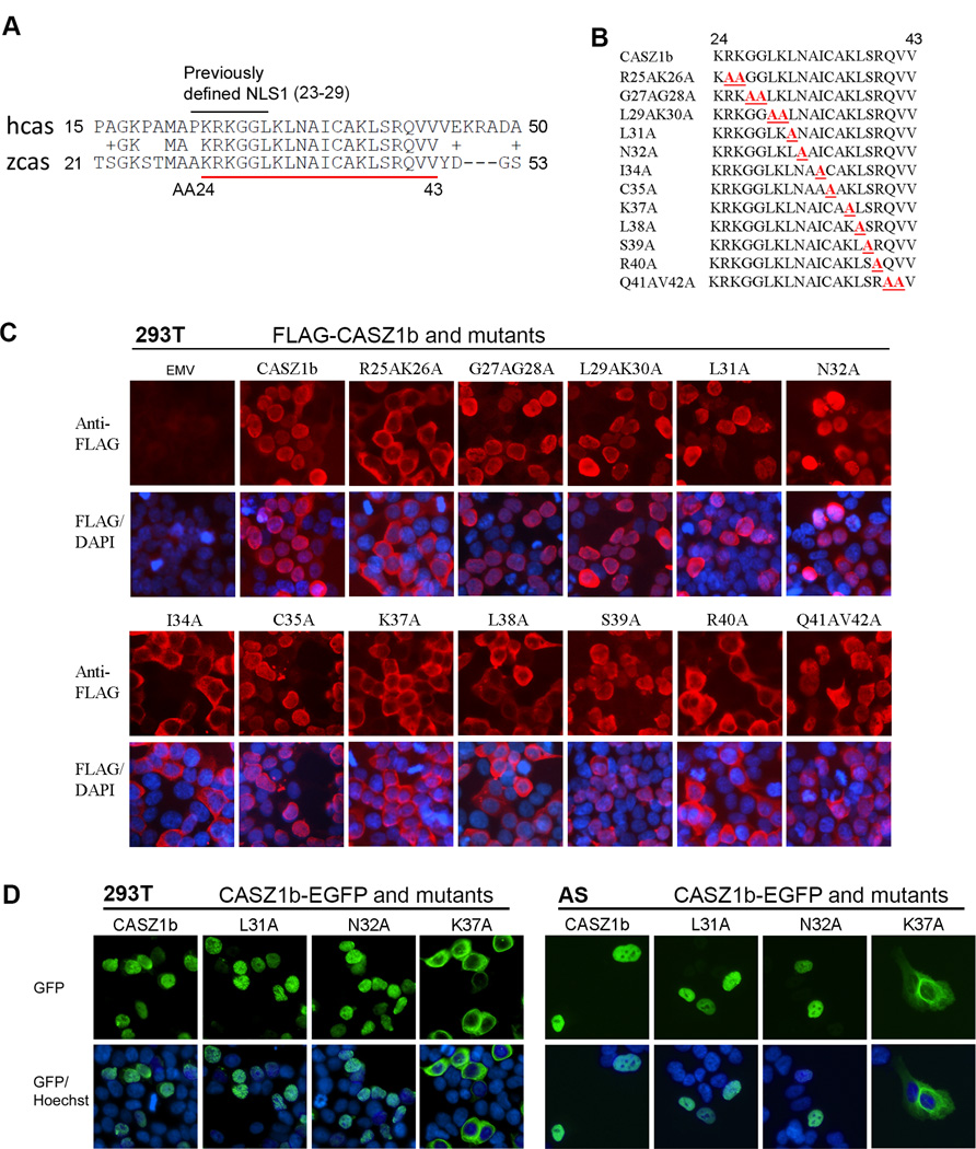
Figure 2. Alanine scanning to characterize previously defined NLS1 and the conserved region close to NLS1.
(A) The alignment of human CASZ1 (hcas) and zebrafish CASZ1 (zcas) at the N-terminus shows that AA23–42 are evolutionarily conserved. The previously defined NLS1 is marked on top and conserved AAs are underlined in red. (B) The alanine scanning mutations generated in the N-terminus conserved region of CASZ1. (C) Assessment of cellular localization of CASZ1b variants. FLAG-tagged CASZ1b WT and N-terminus deletion constructs were transfected into 293T cells and stained with anti-FLAG antibody and DAPI 24 hr after. (D) Assessment of cellular localization of CASZ1b variants. CASZ1b–EGFP and mutant constructs were transiently transfected into 293T cells (top panel) or SK-N-AS cells (bottom panel) overnight, then the cells were stained with Hoechst.

