Figure 7.
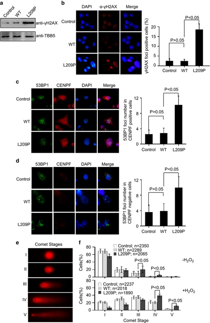
The FEN1 L209P mutation induces spontaneous DNA damage. (a) Western blotting assay of γH2AX in L209P FEN1-containing cells. (b) Representative picture of γH2AX immunofluorescence in L209P FEN1-containing SW480 cells. Red indicates γH2AX and blue indicates DAPI. The right panel shows the percentage of nuclei that contain γH2AX foci. (c and d) Representative picture of 53BP1 and CENPF immunofluorescence in L209P FEN1-containing SW480 cells. Green indicates 53BP1, red indicates CENPF and blue indicates DAPI. The graph in the right panel shows the 53BP1 foci number in the CENPF positive (c) or negative (d) cells. A total of 60 CENPF-positive (G2) or -negative (G1) cells were scored for each cell line. (e) Typical examples of cell nuclei representing the comet stages I–V. (f) Distribution and statistical evaluation of the Comet stages of the L209P FEN1, WT FEN1 or parental SW480 cells. Cell distributions are shown before (up panel) and 24 h after H2O2 treatment (bottom panel). In all panels, values are the mean±s.d. of three independent experiments.

