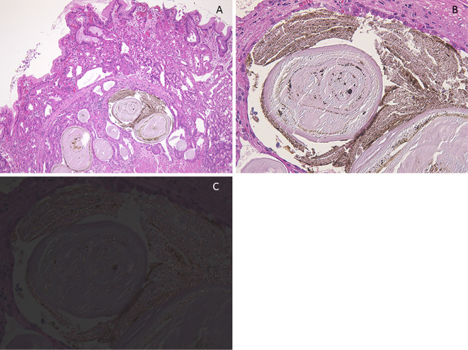
Figure 5.
(Case 5). Histologic findings of black spot (BS). A: Parietal cell protrusions, fundic gland cysts (FGCs) and brownish pigmentation in the FGCs [Hematoxylin and Eosin (H&E) staining, 40×]. B: Amorphous eosinophilic contents and brownish pigmentation in the FGCs (H&E staining, 400×). C: Brownish pigmentation includes tiny birefringent crystals (polarizing observation method, 400×).

