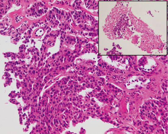
Figure 1.

Biopsied specimens stained with Hematoxylin and Eosin staining show small necrotic foci (inset) and tumor cells with fine nuclear chromatin and moderate eosinophilic cytoplasm growing in a trabecular pattern and organoid nesting arrangement. Only one mitosis was found in 10 high-power fields.
