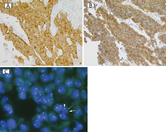
Figure 2.
HC for (A) synaptophysin and (B) ALK. (C) FISH analysis of the ALK locus using a break-apart probe strategy. Strongly positive staining for synaptophysin (Bb) (A) and ALK (5A4, Nichirei Biosciences, Tokyo) (B) was observed in tumor cells. (C) Approximately 74% of the tumor cells showed rearrangement at the ALK locus.

