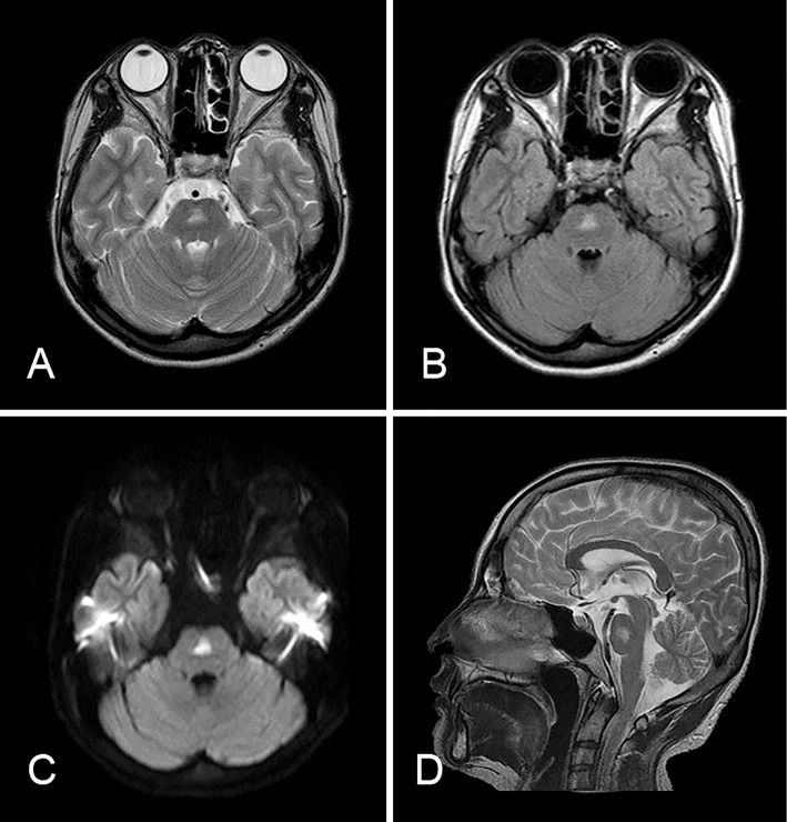
Figure 2.
MRI of the brain in Case 2. A: T2-weighted image. B: FLAIR image. C: A diffusion-weighted image revealed a hyperintense lesion in the pons with slight edema. D: Sagittal T2-weighted imaging of the brainstem showed the lesion existed on the pontine midline. The lesion finally diminished without any treatment.

