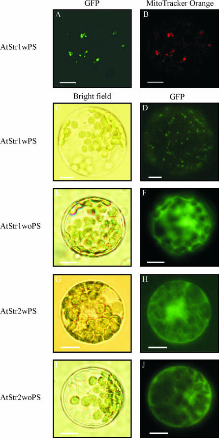
Figure 1.
Subcellular localization of AtStr1 and AtStr2 GFP fusion constructs with and without signal peptide. A, Arabidopsis protoplasts were transformed with AtStr1 including its targeting peptide sequence (AtStr1wPS). Fluorescence images of the protoplasts were taken using a confocal laser scanning microscope. The GFP fluorescence was excited with the argon laser (488 nm) and detected at 515 nm to 520 nm. B, The same protoplast suspension was additionally stained with MitoTracker Orange CMTMRos. The fluorescence was excited with the green helium neon laser (543 nm) and detected at 575 nm to 585 nm. C–J, Arabidopsis protoplasts were transiently transformed with AtStr1 and AtStr2 constructs with and without the targeting peptide sequences (AtStr1wPS/AtStr1woPS and AtStr2wPS/AtStr2woPS, respectively). Bright field images shown in C, E, G, and I were made to visualize the protoplast's cell membrane and the chloroplasts. Fluorescence images of the protoplasts shown in D, F, H, and J were taken using an Axioskop microscope with filter sets optimal for GFP fluorescence (BP 450–490/LP 520). All scale bars represent 10 μm.

